

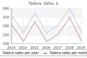
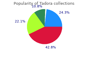
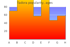
Tadora
"Generic 20mg tadora, does kaiser cover erectile dysfunction drugs".
By: N. Boss, M.A., Ph.D.
Deputy Director, Emory University School of Medicine
Thus a homeopathic top rated erectile dysfunction pills cheap tadora 20 mg visa, vitamin or mineral that might help the patient also might produce a change in the field erectile dysfunction medication free samples discount 20mg tadora fast delivery. Our purpose is to understand this phenomenon through the development of the electrical reactivity work we have done erectile dysfunction 5-htp discount 20 mg tadora. The Xrroid takes in scores of capacitance, inductance, resistance, voltage, amperage, etc. They were discovered from systemic infections, throat cultures, vaginal cultures, blood cultures for fungus (including the entire candida family), cryptococcus and blastomycoses. Forty patients were selected who had presented with symptomatic problems showing a good indication of the infection. The culture was then performed, which then proved undeniably that the infection existed in these thirty-five patients. These patients were then tested by the Xrroid to determine their electrical reactivity to the infectious agents. Results: Correlation between the infection and the Xrroid results came out at ninety-three percent correlation, showing that the Xrroid is indeed accurate in predicting the existence of a pathogenic organism that could be cultured in the human body. These results will help us to further understand the idea of electrical reactivity and medication testing, and the phenomenon of the Xrroid electrical reactivity test. Discussion: As we have seen, the Xrroid was able to pick the electrical reactivity of patients who had pathological levels of an organism. As we have pointed out in other research, the Xrroid also has a strong correlation in mineral analysis, chromosomes, infections (in this study), vitamins, minerals, and other measures of electrical reactivity. We can then find various ways to help him, using simple interventions in a prevention mode, such as homeopathics, nutrition, and lifestyle and behavioral changes. In developing a system of prevention medicine, we need some simple, inexpensive ways to determine whether the patient is at risk of a disease or infection. The Xrroid is very helpful in developing such a system, as it can pick up the initial electrical reactivity sometimes even far before the pathological level of the microorganism is reached. Then the system can properly deal with these items in a more positive fashion, if intervention is early enough. The system of antibiotics that modern medicine has us ed for years is a system of crisis-oriented medicine, in which the organism is not treated until pathogen levels are high. Because of the harshness of synthetic chemicals, which have dramatic side effects, the doctor must be absolutely sure that the level of the microorganism is definitely threatening the patient before he prescribes these chemicals. As an earlier but simpler intervention system, the Xrroid can help provide indication which can gear the doctor towards using a simple homeopathic, vitamin, mineral, or simple behavioral techniques to prevent the pathogen from increasing its numbers. Thus in developing such a system of medicine, we definitely need an early warning device, for which the Xrroid has been designed. Certain patients have high levels of a microorganism that they might have ignored for months or years. In this type of case, if it is extreme and the reticuloendothelial system cannot be pushed, perhaps the use of these harsh antibiotics should be resorted to. Simple homeopathics, as we have shown in our other studies, can be used to help turn these infections around in a quick and easy way. A new type of medicine can be developed based on early warning signs and simple behavioral, nutritional and natural treatment. Many people are addicted to coffee, sugar or cigarettes, or in some cases the harder substances such as cocaine, heroine, barbiturates and amphetamines. There also are addictive behaviors to sex, anger, fear, and other types of life patterns. Alcoholism is yet another extremely addictive behavior pattern that can cause tremendous problems. In dealing with alcoholism and other addictions, one of the finest programs in the world today is the twelvestep program authored by Alcoholic*s Anonymous. For further information and help, we heartily recommend that you seek out Alcoholic*s Anonymous, Gambler*s Anonymous, or whatever agency is applicable for the type of addictive problem your client has. Group therapy can be extremely helpful for clients to overcome addiction and the negative aspects that addiction produces. Your client must first accept the fact that there is a problem before he can seek help to overcome his problem. If the person does not accept that there is a problem, it will be hard for him to get help.
The two thalami are usually connected by a band of grey matter called the interthalamic connexus erectile dysfunction doctor near me purchase tadora 20mg without prescription, which passes through the ventricle impotence young male order tadora 20mg with visa. The lateral wall impotence from steroids cheap tadora 20 mg otc, below the hypothalamic sulcus, is formed by the medial surface of the hypothalamus. Note the mode of formation of the tela choroidea that lies in the roof of the ventricle c. A small part of the lateral wall, above and behind the thalamus, is formed by the epithalamus. The interventricular foramen is seen on the lateral wall, just behind the column of the fornix. The anterior wall of the third ventricle is formed mainly by the lamina terminalis. Its upper part is formed by the anterior commissure, and by the columns of the fornix as they diverge from each other. The roof of the ventricle is formed by the ependyma that stretches across the two thalami (55. Within the tela choroidea, there are two plexuses of blood vessels (one on either side of the middle line) which bulge downwards into the cavity of the third ventricle. The cavity of the third ventricle shows a number of prolongations or recesses (55. The pineal recess lies between the superior and inferior lamina of the stalk of the pineal body. The tela choroidea is a double-layered fold of pia mater that occupies the interval between the splenium of the corpus callosum and fornix, above, and the two thalami below. Its posterior end is broad and lies in the gap between the splenium (above) and the posterior part of the roof of the third ventricle (below) (55. The anterior end (representing the apex of the triangle) lies near the right and left interventricular foramina. Its right and left lateral edges project into the central parts of the corresponding lateral ventricles (55. When traced posteriorly, the two layers of pia mater forming the tela choroidea separate. The choroid plexuses are highly vascular structures that are responsible for the formation of cerebrospinal fuid. The surface of each plexus is lined by a membrane formed by fusion of the ventricular ependyma with the pia mater of the tela choroidea. Microscopic examination shows that the surface of the choroid plexus has numerous villous processes. Each process contains a plexus of capillaries that are connected to afferent and efferent vessels. Because of the presence of these processes, the surface area of the choroid plexuses is considerable. Four choroid plexuses are to be seen in relation to the tela choroidea of the third and lateral ventricles. Two of these (one right and one left) lie along the corresponding lateral margins, and project into the central part of the corresponding lateral ventricle. Two other plexuses run parallel to each other, one on either side of the middle line. At each posterolateral angle of the tela choroidea, the choroid plexus of the lateral ventricle continues into the inferior horn. The tela choroidea and choroid plexuses of the fourth ventricle are considered later in this chapter. For a proper understanding of the anatomy of the fourth ventricle, it is necessary that some features of the gross anatomy of the cerebellum and of related structures be clearly understood. When traced inferiorly (and posteriorly) the velum merges into the white matter of the cerebellum. It will be recalled that the nodule forms the most anterior part of the inferior vermis.
Adjoining part of little fnger geal joint fexor retinaculum Opponens digiti minimi 1 erectile dysfunction cures generic tadora 20mg online. Hook of hamate Medial surface of 5th Flexes the ffth Deep branch of ulnar (distal part) metacarpal bone metacarpal bone nerve (C8 impotence under hindu marriage act order 20 mg tadora, T1) 2 drugs for treating erectile dysfunction order tadora 20 mg with visa. Adjoining part of and rotates it laterally fexor retinaculum (makes palm hollow) 6. Insertion of each muscle into dorsal digital expan- expansion of one digit sion of one digit 4. They fex the metacarpo-phalangeal joint and and extend the interphalangeal joints of the extend the interphalangeal joints of the digit con- digit concerned cerned Contd. A dorsal interosseus muscle is always inserted not be inserted into the base of the proximal into the base of the proximal phalanx of the digit phalanx concerned 5. Dorsal interossei take origin from all fve meta- inserted into the frst, second, fourth, and carpals and are inserted into the second, third and ffth digits (not the third) fourth digits (not frst and ffth) 6. The median nerve is formed by union of lateral and medial roots that arise from the corresponding cords of the brachial plexus. Near the middle of the arm it crosses superfcial to the artery to reach its medial side, and descends in this position to the cubital fossa. The nerve leaves the cubital fossa by passing between the superfcial and deep heads of the pronator teres. It runs down the forearm in the plane between the fexor digitorum superfcialis and the fexor digitorum pro- fundus. At the wrist the nerve lies between the tendons of the fexor digitorum superfcialis (medially) and the fexor carpi radialis (laterally). The pronator teres is supplied by a branch that arises in the lower part of the arm. Direct branches arising in the upper part of the forearm supply the fexor carpi radialis, the palmaris longus and the fexor digitorum superfcialis. The anterior interosseous nerve arises from the median nerve as the latter passes between the two heads of the pronator teres. The muscles supplied through it are the fexor pollicis longus, the lateral part of the fexor digitorum pro- fundus and the pronator quadratus. A muscular branch arising in the palm supplies the thenar muscles namely the fexor pollicis brevis, the abduc- tor pollicis brevis and the opponens pollicis. The frst and second lumbrical muscles of the hand are supplied by branches from the digital nerves. The palmar cutaneous branch (superfcial palmar branch) arises in the lower part of the forearm, and passes into the hand superfcial to the fexor retinaculum. The median nerve ends by dividing into a variable number of palmar digital branches that subdivide so that ultimately seven proper palmar digital nerves are formed: two each (one medial and one lateral) for the thumb, the index and the middle fngers, and one for the lateral half of the ring fnger. Through these branches the median nerve supplies the palmar surface of the lateral three and a half digits. It also supplies the dorsal surfaces of the terminal parts of the same digits including the nail beds, the skin over the terminal phalanx of the thumb, and over the middle and terminal phalanges of the index and mid- dle fngers and the lateral half of the ring fnger. Articular branches arising directly from the median nerve near the elbow supply the elbow joint and the superior radioulnar joint 2. The distal radioulnar joint and the wrist joint are supplied through the anterior interosseous nerve. The metacarpophalangeal and interphalangeal joints are supplied through the digital branches. CliniCal Correlation Effects of Injury to the Median Nerve the effects of injury to the median nerve vary depending upon the level of injury, the effects being confned to structures supplied by branches distal to the injury. Unopposed action of the fexor carpi ul- naris adducts the hand when fexion is attempted.
Uterine ?broids can often be palpated on examination of the the intravenous pyelogram in Figure 4 erectile dysfunction injection therapy 20mg tadora. Their identity can contrast throughout a normal collecting system erectile dysfunction band order 20mg tadora, outlining easily be established by bedside ultrasonography erectile dysfunction treatment options articles buy discount tadora 20mg on line. Multiple showing another case of multiple calci?ed ?broids extending calci?ed phleboliths in the pelvis are incidentally noted, out of the pelvis (white arrows). Plain radiography is not considered su?cient to exclude ureterolithiasis because most ureteral stones, although Renal stone radiopaque, are too small to be identi?ed on plain ?lm or the patient in Figures 4. She had had several previous episodes of case, demonstrating the di?culty of identifying ureteral 10:28:08 04 Chapter 4: Plain Film Evaluation of the Abdomen Figure 4. This spontaneously, and the initial management of all but the stone was not identi?ed with certainty but was con?rmed largest stones is expectant anyway. Pancreatic calci?cations This image shows the characteristic stippling of pancreatic calci?cations. They extend from the right upper quadrant, across the midline, to the left upper quadrant (black arrows). A follow-up study and surgical obtained in a patient complaining of low back pain. There are an estimated 11,000 new cases of patients, soft tissue often obscures the C7-T1 junction. The vertebral bodies, 2% to 6% of all blunt trauma patients and may be present in interbody spaces, transverse processes, articular masses with up to one-third of those who present unconscious (1, 2). The open-mouth odontoid view Most C-spine injuries are the result of blunt trauma from should show the articulation of the lateral masses of C1 and motor vehicle collisions, falls, sports-related injuries, and C2 and the entire dens. Decades of biomechanical studies have correlated Flexion-extension (F/E) radiographs. Signs of instability on F/E imaging regions and patterns of injury within the C-spine. One-third of C-spine fractures occur at C2, and about one-half between C5 and C6 (3). Noncontiguous injuries are common, espe- cially in patients with severe mechanism of injury. The same study, and many others, found an increased incidence of C-spine Ruptured transverse ligament of C1 Least fractures with increasing Injury Severity Score. The most widely accepted classi?cation ligamentous disruption of C-spine injury stability was described by Trafton Hyperextension fracture-dislocation (Table 5. Compression fracture of C2 with anterior or Plain radiography of the C-spine was the traditional posterior displacement (hangman fracture) screening test of choice in patients with suspected C-spine Extension teardrop fracture injury. This displacement (Je?erson fracture) controversy and its supporting data and practice guidelines Unilateral facet dislocation will be covered later in this chapter. Step one is to assess for any high-risk factor that would immediately mandate include >3. Step two is to assess for any low-risk factor that cent disks, displaced apophyseal joints, widened disk spaces, will allow for active range-of-motion testing. Step three is loss of >30% of disk height, or presence of prevertebral to test active range of motion by asking patients to actively hematoma, as evidenced by abnormal thickness or contour rotate their necks 45 degrees to both sides. In its derivation study, the rule was 100% sensi- injury, and F/E imaging has not been shown to add signi?- tive and 42. In addition, the four missed injuries in the performed on obtunded patients or those who have pain study were categorized as not clinically signi?cant. The criteria were ing the indications for radiologic screening for C-spine injury validated in a prospective study of 34,069 patients, in in blunt trauma patients. In obtunded patients with ries by the criteria had clinically signi?cant injuries.
When z verse magnetization Mxy and to regain equilibrium is 1 t T1 osbon erectile dysfunction pump order cheapest tadora and tadora, Mz is (1 e ) 63 the longitudinal relaxation time but since this is an 2 t 2 T1 erectile dysfunction dsm 5 order tadora 20mg visa, Mz is (1 e ) 86 exponential event the time constant itself is meas- 3 t 3 T1 impotence urology order 20 mg tadora free shipping, Mz is (1 e ) 95 ured when the longitudinal magnetization reaches 4 t 4 T1, Mz is (1 e ) 98 63% of its original value. Local deviations in the micro- Liver 350 420 500 40 scopic magnetic fields generated by interactions Kidney 430 590 690 58 between magnetic moments of atoms lead to slight Muscle 550 730 870 45 differences in resonance frequencies which cause Heart 570 750 880 57 phase interference or dephasing of the spins. The composite signals in (b) dephase as the phase During longitudinal relaxation the protons lose diagrams in Fig. With ured here suffers from distortions due to the main simple molecular structures (water), the energy loss magnetic field inhomogeneities and is distinguished by is slower and T1 is long. The exception to this rule is calling it T2* (T2 star, the * denotes a distorted signal fat which, in spite of being a simple compound, has influenced by magnetic field inhomogeneities). The chemical bonds at the ends of its fatty acid molecules additional effect of these inhomogeneities causes Mxy which have frequencies near the Larmor frequency of to decay far more quickly than the expected T2 so hydrogen. Image contrast is a function of T2 in spin quently T1 times are short for fatty tissue. More com- echo images but T2* determines image contrast in plex or solid tissue (muscle protein) absorbs proton fast sequences or gradient echo images. T1 is signal is complex and must be obtained by indirect strongly related to tissue water content; time periods means, to be described later. During equilib- long T2; it follows an exponential slope whose time rium recovery precession of the transverse magneti- constant T2* is taken at 37% maximum. Group 1 consists of small molecules; group 2 consists of 0 1 2 more complex molecules including protein and lipids. Magnet inhomo- geneities obscure the true T2 tissue so the true value of T2 must be separated from magnet inhomogeneities (c) by using a special pulse sequence (described later). All molecules in various values of T2 the calculation of M is as group 2 give short T2 values. The loss of trans- verse magnetization Mxy, given by T2, occurs relatively Figure 19. These can be broadly undergoes relaxation, to gain equilibrium, moving divided into two groups. These the phase relationships lost during the saturation are partial saturation pulse sequences. In an imaging system, where short acquisition times are most important, exact measurement is never attempted. A description of the full T1 and T2 measurement is (a) included here for reference. Total recovery will be given by tissues with a gauss, G: unit of magnetic flux in the c. This longitudinal relaxation: return of Mz to M0 after remains constant until an external force changes excitation. Requires an exchange of energy between the direction of rotation causing precession. Measured nuclei possess intrinsic angular momentum called spin by the time constant T1 B : the magnetic field, measured in tesla magnetic moment, m: given by a nucleus (proton) 0 B : the radio frequency magnetic induction field with spin.
Generic 20 mg tadora with mastercard. Erectile Dysfunction From Diabetes.