

Chloromycetin
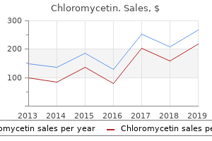
"Chloromycetin 250mg discount, symptoms leukemia".
By: A. Mitch, M.A., M.D., Ph.D.
Deputy Director, Albany Medical College
The third part (4in (10cm)) runs transversely to the left symptoms for mono order chloromycetin paypal, crossing the inferior vena cava medications ranitidine buy chloromycetin cheap online, the aorta and the third lumbar vertebra symptoms gastritis order chloromycetin in united states online. It is surprisingly easy for the surgeon to confuse this with the ileocaecal junction, a mistake which may be disas- trous. He confirms the identity of the duodenal termination by the presence of the suspensory ligament of Treitz, which is a well-marked peritoneal fold descending from the right crus of the diaphragm to the duodenal termina- tion, and by visualizing the inferior mesenteric vein which descends from behind the pancreas immediately to the left of the duodenojejunal junction. The gastrointestinal tract 77 Blood supply of the duodenum The superior pancreaticoduodenal artery arises from the gastroduodenal artery; the inferior pacreaticoduodenal artery originates as the first branch of the superior mesenteric artery. These vessels both lie in the curve between the duodenum and the head of the pancreas, supplying both struc- tures. Interestingly, their anastomosis represents the site of junction of the fore-gut (supplied by the coeliac artery), and the mid-gut (supplied by the superior mesenteric artery), at the level of the duodenal papilla (see page 86). Clinical features 1The first part of the duodenum is overlapped by the liver and gall- bladder, either of which may become adherent to, or even ulcerated by, a duodenal ulcer. The gallstone may then impact in the lower ileum as it traverses the gut to produce intestinal obstruction (gall- stone ileus). Simi- larly, the right kidney lies directly behind this part of the duodenum, which may be injured in performing a right nephrectomy. Within a few minutes of swallowing a barium meal, the first part of the duodenum becomes visible as a triangular shadow termed theduodenal cap. It is in this region that the great majority of duodenal ulcers occur; an actual ulcer crater may be visual- ized, filled with barium, or deformity of the cap, produced by scar tissue, may be evident. The rest of the duodenum can also be seen, the shadow being floccular due to the rugose arrangement of the mucosa. Small intestine The length of the small intestine varies from 10 to 33 feet (3–10m) in 78 The abdomen and pelvis different subjects; the average is some 24 feet (6. Resection of up to one- third or even half of the small intestine is compatible with a perfectly normal life, and survival has been reported with only 18in (45cm) of small intestine preserved. The mesentery of the small intestine has a 6in (15cm) origin from the posterior abdominal wall, which commences at the duodenojejunal junc- tion to the left of the 2nd lumbar vertebra, and passes obliquely down- wards to the right sacro-iliac joint; it contains the superior mesenteric vessels, the lymph nodes draining the small gut and autonomic nerve fibres. The bowel does, however, change its character from above downwards, the following points enabling the surgeon to determine the level of a loop of small intestine at operation. The ileum is supplied by shorter and more numerous terminal vessels arising from complete series of three, four or even five arcades (Fig. Large intestine The large intestine is subdivided, for descriptive purposes, into: caecum with the appendix vermiformis; ascending colon (5–8in (12–20cm)); hepatic flexure; transverse colon (18in (45cm)); Fig. The gastrointestinal tract 79 splenic flexure; descending colon (9–12in (22–30cm)); sigmoid colon (5–30in (12–75cm), average 15in (37cm)); rectum (5in (12cm)); anal canal (1. The large bowel may vary considerably in length in different subjects; the average is approximately 5 feet (1. The colon (but not the appendix, caecum or rectum), bears characteris- tic fat-filled peritoneal tags called appendices epiploicae scattered over its surface. These are three flattened bands commencing at the base of the appendix and running the length of the large intestine to end at the rec- tosigmoid junction. They represent the great bulk of the longitudinal muscle of the large bowel; because the taeniae are about a foot shorter than the gut to which they are attached, the colon becomes condensed into its typical sacculated shape. These sacculations may be seen in a plain radi- ograph of the abdomen when the large bowel is distended and appear as incomplete septa projecting into the gas shadow. The radiograph of dis- tended small intestine, in contrast, characteristically has complete trans- verse lines across the bowel shadow due to the transverse mucosal folds of the valvulae conniventes. Peritoneal attachments The transverse colon and sigmoid are completely peritonealized (the former being readily identified by its attachment to the greater omentum). The ascending and descending colon have no mesocolon but adhere directly to the posterior abdominal wall (although exceptionally the ascending colon has a mesocolon). The caecum may or may not be com- pletely peritonealized, and the appendix, although usually free within its own mesentery, occasionally lies extraperitoneally behind caecum and ascending colon or adheres to the posterior wall of these structures. The rectum is extraperitoneal on its posterior aspect in its upper third, posteriorly and laterally in its middle third and completely in its lower third as it sinks below the pelvic peritoneum. In the fetus it is a direct outpouching of the caecum, but differential overgrowth of the lateral caecal wall results in its medial displacement. The appendix is usually quite free in this position although occasionally it lies beneath the peritoneal covering of the caecum. If the appendix is very long, it may actually extend behind the ascending 80 The abdomen and pelvis Fig.
There is no food effect on absorption symptoms zika virus discount chloromycetin 250 mg otc, minimal (15%) plasma protein binding medicine jar order genuine chloromycetin, and only moderate (20–50%) metabolism; no active metabolites are formed symptoms zoloft overdose buy cheap chloromycetin 250 mg line. Although increased levels are seen with renal failure and hepatic impairment, there is no age or gender effect, no autoinduction, no inhibition of metabolism, and kinetics are linear. Drug interactions do occur and can be complex, but the major effect is on topiramate levels rather than on the levels of other antiseizure drugs. Birth control pills may be less effective in the presence of topiramate, and higher estrogen doses may be required. Vigabatrin is marketed as a racemate; the S(+) enantiomer is active and the R(−) enantiomer appears to be inactive. The half-life is approximately 6–8 hours, but considerable evidence suggests that the pharmacodynamic activity of the drug is more prolonged and not well correlated with the plasma half-life. In adults, vigabatrin should be started at an oral dosage of 500 mg twice daily; a total of 2–3 g (rarely more) daily may be required for full effectiveness. Less common but more troublesome adverse reactions are agitation, confusion, and psychosis; preexisting mental illness is a relative contraindication. The drug was delayed in its worldwide introduction by the appearance in rats and dogs of a reversible intramyelinic edema. This phenomenon has now been detected in infants taking the drug; the clinical significance is unknown. In addition, long-term therapy with vigabatrin has been associated with development of peripheral visual field defects in 30–50% of patients. Newer techniques such as optical coherence tomography may better define the defect, which has proved difficult to quantify. Vigabatrin is usually reserved for use in patients with infantile spasms or with complex partial seizures refractory to other treatments. Its primary site of action appears to be the Na channel; it also acts on T-type 2+ voltage-gated Ca channels. The drug is effective against partial and generalized tonic-clonic seizures and may also be useful against infantile spasms and certain myoclonias. It has good bioavailability, linear kinetics, low protein-binding, renal excretion, and a half-life of 1–3 days. Ethosuximide has very little activity against maximal electroshock but considerable efficacy against pentylenetetrazol seizures; it was introduced as a “pure petit mal” drug. Chemistry Ethosuximide is the last antiseizure drug to be marketed whose origin is in the cyclic ureide structure. Methsuximide and phensuximide have phenyl substituents, whereas ethosuximide is 2-ethyl-2-methylsuccinimide. Mechanism of Action 2+ Ethosuximide has an important effect on Ca currents, reducing the low-threshold (T-type) current. The T-type Ca currents are thought to provide a pacemaker current in thalamic neurons responsible for generating the rhythmic cortical discharge of an absence attack. Inhibition of this current could therefore account for the specific therapeutic action of ethosuximide. Clinical Uses As predicted from its activity in laboratory models, ethosuximide is particularly effective against absence seizures, but has a very narrow spectrum of clinical activity. Documentation of its effectiveness in human absence seizures was achieved with long-term electroencephalographic recording techniques. Data continue to show that ethosuximide and valproate are the drugs of choice for absence seizures and are more effective than lamotrigine. This corresponds to a half-life of approximately 40 hours, although values from 18 to 72 hours have been reported. Therapeutic Levels & Dosage Therapeutic levels of 60–100 mcg/mL can be achieved in adults with dosages of 750–1500 mg/d, although lower or higher dosages and blood levels (up to 125 mcg/mL) may be necessary and tolerated in some patients. The drug might be administered as a single daily dose were it not for its adverse gastrointestinal effects; twice-a-day dosage is common.
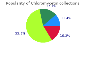
Purchase chloromycetin 500 mg without a prescription. Total Health: HIV - AIDS - Symptoms Facts Prevention & Treatment (Part 1).
The caudal and lateral aspects are traversed by fibers of inferior collicular nuclei show a definite tonotopic the superior cerebellar peduncle treatment laryngitis buy chloromycetin online. The corticorubral projections originate from the precentral and the premotor cortex and project so- matotopically onto the red nucleus medicine 5443 cheap chloromycetin 250mg otc. Fibers originating from the deep cerebel- longitudinal fasciculus; 6 medicine hat jobs discount 250mg chloromycetin, central tegmental tract; 7, medial lar nuclei decussate in the caudal midbrain before lemniscus; 8, spinothalamic tracts; 9, lateral lemniscus; 10, decussation of superior cerebellar peduncle (brachium traversing and surrounding the contralateral red conjunctivum); 11, midbrain reticular formation; 12, interpe- nucleus. Those originating in the dentate nucleus duncular nucleus; 13, substantia nigra; 14, cerebral peduncle; terminate in the rostral third of the contralateral red 15, frontopontine tract; 16, pyramidal tract; 17, nucleus while those from the globose and embo- occipitotemporopontine tract; 18, oculomotor nerve; 19, liform nuclei terminate somatotopically in the cau- brachium pontis; 20, interpeduncular cistern; 21, basilar ar- tery; 22, posterior cerebral artery; 23, lateromesencephalic dal two thirds. Cells of the caudal portion give rise to cistern; 24, ambient cistern the crossed rubrospinal tract, which influences flex- or motor tone. Stimulation of the red nucleus in animals produces flexion of the ipsilateral limb due to the fact that both systems, the superior cerebellar peduncle as well as the rubrospinal tract, are crossed. Clinically, lesions involving the red nucleus are re- sponsible for an ipsilateral oculomotor disturbance associated with contralateral involuntary move- The Brainstem and Cerebellum 233 Fig. This c The Substantia Nigra: Morphology pigmentation is maximum in humans, appearing after and Functional Anatomy the fourth or fifth year of life and increasing in melanin The substantia nigra, also called the locus niger, is locat- content with age. The neurons of the pars compacta ed in the midbrain between the crus cerebri and the contain high concentrations of dopamine whereas the tegmentum, as shown on the parasagittal (Figs. Efferent connections of the substantia nigra are represented by the nigrostriatal dopaminergic fibers arising from the pars compacta and projecting to the striatum, the rostral two thirds of the substantia ni- gra terminating in the head of the caudate nucleus and the caudal part in the putamen. A reciprocal ar- rangement characterizes the striatonigral and the nigrostriatal fibers constituting a closed feed-back loop. In pri- mates, the nigrothalamic fibers terminate in those thalamic motor nuclei lacking input from the cere- bellum or the basal ganglia. The nigrotectal fibers A end in the superior colliculus and play an important role in the initiation of saccadic eye movements (Hi- kosaka and Wurtz 1983a–d). The nigrotegmental fi- bers terminate in the pedunculopontine nucleus, the latter projecting fibers back to the pars compacta of the substantia nigra, constituting a nigrotegmenton- igral feed-back loop. The importance of the substantia nigra is due to its involvement in many diseases of the basal ganglia, such as Parkinson’s disease and other parkinsonian syndromes of different etiologies. Most of these dis- eases are characterized by involuntary movements such as tremor, dystonia, chorea, athetosis, or bal- lism. A,B 1, Quadrigeminal plate; 2, central gray matter; logic basis of this idiopathic disease is a massive loss 3, lateral lemniscus; 4, medial lemniscus; 5, substantia nigra; of the pigmented cells in the substantia nigra and the 6, cerebral peduncle (crus cerebri); 7, subthalamic nucleus; 8, ventral tegmental area. The nigral cell loss ranging thalamic fasciculus; 9, zona incerta; 10, Forel’s fields H; 11, from 50% to 90% and is associated with reactive gli- centromedian nucleus of thalamus; 12, pulvinar thalami; 13, osis and the presence of Lewy bodies. Degeneration ventral anterior and lateral nuclei of thalamus; 14, lateral posterior thalamic nucleus; 15, internal medullary lamina; 16, of the nigral neurons is responsible for the marked optic tract; 17, globus pallidus; 18, putamen; 19, caudate decrease in dopamine concentration in the striatum, nucleus; 20, anterior commissure; 21, genu of internal cap- the dopaminergic depletion being more significant sule; 22, splenium of corpus callosum; 23, isthmus; 24, tento- in the putamen than in the caudate nucleus and the rium cerebelli; 25, superior surface of the cerebellar hemi- nigrostriatal system being more severely affected sphere; 26, inferior surface of cerebellar hemisphere; 27, den- tate nucleus; 28, inferior cerebellar peduncle; 29, pontine nu- than the mesolimbicocortical system. The decrease clei; 30, intracavernous carotid artery; 31, intracanalicular in striatal dopamine is directly proportional to the optic nerve; 32, gyrus rectus neuronal degeneration in the substantia nigra. On coronal cuts, the crus cerebri constitute the me- dial boundary of the lateral wings of the transverse cerebral fissure (Figs. The exact topography of these corticofugal fibers in the crus cerebri differs to some extent according to different authors. Déjerine (1901) places the corti- cospinal and the corticobulbar fibers in the medial three fifths of the cerebral peduncle and considers that they are somatotopically arranged. Classically, the and ventral trigeminothalamic tract; 8, lateral and ventral corticospinal and corticobulbar fibers occupy the spinothalamic and spinotectal tracts; 9, lateral lemniscus; 10, middle two thirds of the crus cerebri, that is, its ex- decussation of brachium conjunctivum; 11, brachium pontis; 12, frontopontine tract; 13, pyramidal tract; 14, treme medial and lateral portions, containing corti- occipitotemporopontine tract; 15, pontocerebellar fiber copontine fibers, the frontopontine fibers located bundle; 16, basilar artery; 17, prepontine cistern; 18, medially and the temporopontine, parietopontine intracavernous and internal carotid artery; 19, culmen and occipitopontine fibers located laterally. The cor- cerebelli; 20, superior edge of tentorium cerebelli (delimiting ticospinal fibers are topographically arranged, the fi- foramen ovale of Pacchioni) bers concerned with the upper extremity being dis- posed more laterally than the most medial fibers 1 Isthmus Rhombencephali: Upper Pons Level innervating the facial musculature. The medial and lateral portions correspond each to a sixth of the This transverse cut orthogonal to the brainstem long crus cerebri. This cut, rostral to the cerebellum The pons derives from the mesencephalic vesicle and immediately caudal to the mesencephalon, is constituting the anterior portion of the hindbrain. It bordered laterally by the inferior medial aspects of may be subdivided into a dorsal portion, the tegmen- the temporal lobes in a horizontal orientation of the tum, and a ventral portion, the pons proper, which is cuts. As completed for be divided into a posterior or roof portion, a tegmen- the midbrain, and in order to facilitate imaging-clin- tal portion anterior to the latter and a ventral portion.
For more than 100 years medicine 101 generic chloromycetin 250 mg online, it has been extracted and used in clinical medicine symptoms questionnaire generic chloromycetin 250 mg overnight delivery, mainly as a local anesthetic and to dilate pupils in ophthalmology treatment yeast infection men order 500 mg chloromycetin otc. Sigmund Freud famously proposed its use to treat depression and alcohol dependence, but addiction quickly brought an end to this idea. Cocaine hydrochloride is a water-soluble salt that can be injected or absorbed by any mucosal membrane (eg, nasal snorting). When heated in an alkaline solution, it is transformed into the free base, “crack cocaine,” which can then be smoked. Inhaled crack cocaine is rapidly absorbed in the lungs and penetrates swiftly into the brain, producing an almost instantaneous “rush. In the central nervous system, cocaine blocks the uptake of dopamine, noradrenaline, and serotonin through their respective transporters. Cocaine exposure increases the risk for intracranial hemorrhage, ischemic stroke, myocardial infarction, and seizures. The term “crack-baby” was used to describe a specific syndrome of the newborn, and the mothers faced harsh legal consequences. The follow-up of the children, now adults, does not confirm a drug-specific handicap in cognitive performance. Moreover, in this population the percentage of drug-users is comparable to controls matched for socioeconomic environment. Tolerance may develop, but in some users a reverse tolerance is observed; that is, they become sensitized to small doses of cocaine. Amphetamine, methamphetamine, and their many derivatives exert their effects by reversing the action of biogenic amine transporters at the plasma membrane. Normal vesicular release of dopamine consequently decreases (because synaptic vesicles contain less transmitter), whereas nonvesicular release increases. They are often produced in small clandestine laboratories, which makes their precise chemical identification difficult. They differ from ecstasy chiefly in the context of use: intravenous administration and “hard-core” addiction is far more common with amphetamines, especially methamphetamine. In general, amphetamines lead to elevated catecholamine levels that increase arousal and reduce sleep, whereas the effects on the dopamine system mediate euphoria but may also cause abnormal movements and precipitate psychotic episodes. Effects on serotonin transmission may play a role in the hallucinogenic and anorexigenic functions as well as in the hyperthermia often caused by amphetamines. Amphetamines are typically taken initially in pill form by abusers, but can also be smoked or injected. Within hours after oral ingestion, amphetamines increase alertness and cause euphoria, agitation, and confusion. Effects on heart rate may be minimal with some compounds (eg, methamphetamine), but with increasing dosage these agents often lead to tachycardia and dysrhythmias. This is perhaps not surprising, because the main effect of ecstasy appears to be to foster feelings of intimacy and empathy without impairing intellectual capacities. This release is so profound that there is a marked intracellular depletion for 24 hours after a single dose. With repetitive administration, serotonin depletion may become permanent, which has triggered a debate on its neurotoxicity. Other complications include serotonin syndrome (mental status change, autonomic hyperactivity, and neuromuscular abnormalities, see Chapter 16) and seizures. This is particularly true in the case of a massive overdose, in which reversal of drug action may be a life- saving measure. Pharmacologic interventions may also aim to alleviate the withdrawal syndrome, particularly after opioid exposure. On the assumption that withdrawal reflects at least in part a hyperactivity of central adrenergic systems, the α -adrenoceptor2 agonist clonidine (also used as a centrally active antihypertensive drug, see Chapter 11) has been used with some success to attenuate withdrawal. Today, most clinicians prefer to manage opioid withdrawal by very slowly tapering the administration of long-acting opioids. Another widely accepted treatment is substitution of a legally available agonist that acts at the same receptor as the abused drug.