Erectafil
"20 mg erectafil otc, erectile dysfunction treatment dallas texas".
By: P. Cyrus, MD
Co-Director, University of Michigan Medical School
The imaging coil should be chosen to maximize the signal-to-noise ratio over the entire body region to be examined erectile dysfunction caused by hernia generic 20mg erectafil visa. Adult head or knee coils are often appropriate for infants weighing less than 10 kg erectile dysfunction protocol scam or not generic erectafil 20mg amex, and adult cardiac coil for medium-sized children weighing between 10 and 40 kg erectile dysfunction natural herbs generic 20mg erectafil fast delivery. More recently, multichannel coils designed specifically for cardiovascular imaging in infants and young children have become commercially available (Fig. Adequate coil coverage and placement should be confirmed early in the examination by reviewing the localizing images. Safety Standard clinical imaging scanners present no known hazards to biologic materials. Animal studies evaluating the influence of static magnetic fields have not demonstrated significant biologic effects for fields of up to 2 T (19). Subsequent investigations found that patients with renal failure were at risk for developing this rare but severe complication (26,27). Consequently, most institutions have adopted a policy to avoid using Gd contrast in patients with impaired renal function (28). Fortunately, surgical clips and sternotomy wires implanted in the chest and abdomen are typically only weakly ferromagnetic. A: Eight- channel knee coil used in infants weighing 4 kg or less; B: Eight-channel pediatric coil used in patients weighing between 4 and 10 kg; C: Five-channel cardiac coil used in patients weighing between 10 and 30 kg; and D: Thirty-two–channel cardiac coil used in patients weighing 30 kg or more. These body weights are approximates and other considerations are taken into account when selecting a coil for an individual patient. The unpredictable nature of the anatomy and hemodynamics often require adjustment of the examination protocol, modification of imaging planes, changing sequences, and adjustment of imaging parameters. Reliance on standardized protocols and postexamination review alone in these patients may result in incomplete or even erroneous interpretation. In practice, the anatomy in question is often assessed by more than one imaging sequence, yielding overlapping information that increases diagnostic confidence. In clinical practice, such inhomogeneities are mostly owing to ferromagnetic implants such as sternal wires, prosthetic heart valves, stents, coils, or other implants. As a result, image acquisition can be completed during 10 to 15 seconds of breath holding. The sequence is repeated N2-D times, each with a different phase- encoding value, until all lines of k-space are collected and a 2-D image is reconstructed. Examples of clinical applications include assessment of tissue characteristics (e. The signal from stationary or relatively slow-moving tissues (such as the myocardium) is gray because the spins within the selected slice have reduced signal intensity (i. On the other hand, blood that flows into the slice contains unsaturated spins that produce relatively strong signals, hence the term “bright blood” imaging (Fig. An important feature of gradient echo sequences is high imaging speed, which allows reconstruction of multiple images during the cardiac cycle that can be displayed in cine loop format. A: T1-weighted image showing a slightly hyperintense globular tumor (arrow); B: T2-weighted image showing the tumor is markedly hyperintense, consistent with a vascular tumor. The brightness of a given tissue in this case is determined primarily by the ratio of T2-to-T1 relaxation times which results in high contrast between the blood pool (T2/T1 = 360 ms/1,200 ms = 0. The motion of the diaphragm is tracked with a navigator pulse and image data are accepted only when the position of the diaphragm lies within a narrow window defined by the operator (Fig. The volume elements (voxels) of the image dataset are typically isotropic, which allows offline reformatting of the image volume in any arbitrary plane. This imaging technique does not require administration of contrast agents and provides high-resolution static imaging of both intracardiac and extracardiac anatomy (Fig. This technique has been used successfully in patients with various congenital anomalies of the coronary arteries (Fig. Examples of common clinical applications include imaging of the aorta and its branches, pulmonary arteries, pulmonary veins, systemic veins, aortopulmonary and venous-venous collateral vessels, systemic-to-pulmonary artery shunts, conduits, and vascular grafts (11,46).


However erectile dysfunction treatment without medicine buy generic erectafil 20 mg, the fnal height of an individual is determined by his/her genetic potential erectile dysfunction pills free trial order erectafil in united states online. The growth plate erectile dysfunction doctors in charleston sc purchase generic erectafil online, also known as physis, is present between the epiphysis and metaphysis at the ends of long bones. It comprises of fve zones: resting zone, proliferative zone, hypertrophic zone, calcifcation zone, and ossifcation zone, 1 Disorders of Growth and Development: Clinical Perspectives 11 from epiphysis to metaphysis. The process of linear growth initiates at the epiphy- seal end of growth plate and new bone is laid down at the metaphysis (Fig. Linear growth is a result of a well-regulated and coordinated process called “chondro-osteogenesis,” which includes chondrocyte proliferation, differenti- ation/hypertrophy, apoptosis, and endochondral ossifcation. Longitudinal bone growth occurs at the epiphyseal growth plate located at the ends of long bones. Later, the growth plate is invaded by blood vessels and bone cell precursors from metaph- ysis, resulting in remodeling of cartilage into bone, a process termed as endo- chondral ossifcation. The various endocrine and paracrine factors that regulate “chondro-osteogenesis” are depicted in the fgure given below (Fig. The linear bone growth occurs at epiphyseal growth plate (at the end of long bones), while circumferential bone growth (appositional bone growth) occurs beneath the periosteum at diaphysis. The appositional bone growth is the result of intramembranous ossifcation, where osteoblast forms the new bone just beneath the periosteum. Periosteal new bone formation is accompanied with endosteal bone resorption as the new bone formation exceeds bone resorption at periosteum and vice versa at endosteum, thereby resulting in increased circumfer- ential bone growth (Fig. In addition, these hormones also have a direct effect on epiphyseal growth plate and promote chondrocyte proliferation. Boys are taller than girls because of physiological delay in the initiation of puberty by a period of 2 years (thereby yielding two additional years of cumula- tive linear growth), more intense pubertal growth spurt, and presence of growth- promoting genes on Y(Yq) chromosome. This difference is due to growth accu- mulated during two additional prepubertal years (10 cm) and the greater gain in 14 1 Disorders of Growth and Development: Clinical Perspectives height during pubertal growth spurt (3 cm) in boys. This knowledge is impor- tant and is used in the calculation of midparental height of an individual. In addition, there is an increase in leptin levels in obese children, which also acts as skeletal growth factor. Further, increased aromatization of androgens to estrogens as a result of excess adiposity also contributes to the linear growth. However, the fnal adult height in obese children does not differ from nonobese children, as a result of early puberty and excess aromatization of androgens leading to prema- ture epiphyseal closure (Fig. Therefore, presence of short stature in an obese child is almost always patho- logical and should be evaluated further. The common causes of short stature with obesity include Cushing’s syndrome, hypothyroidism, isolated growth hormone defciency, pseudohypoparathyroidism, and Prader–Willi syndrome. Thyroxine is responsible for facial bone growth and maturation during prenatal and infantile period. Infants with congenital hypothyroidism therefore have characteristic facial fea- tures including immature facies, flat nasal bridge, and pseudohyper- telorism. Therefore, patients with congenital growth hormone deficiency manifest with frontal bossing, midfacial hypoplasia, and micrognathia. During peripubertal period, gonadal steroids play an impor- tant role in facial maturation and lead to sexual dimorphism in the facial characteristics (Fig. Measurement of body proportions helps in the differential diagnosis of short stature. The presence of body proportions which are disproportionate to the chronological age defnes disproportionate short stature. The causes include hypothyroidism, rickets, skeletal dysplasias, and mucopolysaccharidosis (Fig. What is the importance of measurement of parental height in a child with short stature? Genetic factors have a signifcant contribution to the fnal adult height of an individual. Therefore, an estimate of the genetic potential for the fnal adult height of an individual can be predicted on the basis of height of the parents. Children of short parents are not as short as their parents, and, similarly, chil- dren of tall parents are not as tall as their parents due to the phenomenon of regression to the mean. Therefore, the concept of target height was introduced to predict the fnal adult height of an individual with allowance for regression to the mean.

This condition is known as disc the external and internal ends of the Müller sup- edema erectile dysfunction treatment with fruits erectafil 20 mg without a prescription, papilledema erectile dysfunction age onset proven 20 mg erectafil, or choked disc and can porting cells impotence pronunciation cost of erectafil, the modifed glial cells of the retina. Two parts of the retina that are structurally and functionally different from the rest of the retina are the central area and the optic disc. Embryologically, the retina develops from The central area contains the macula lutea and the diencephalon; hence, it is a central nervous the fovea centralis. The fovea is the area for acute vision, nerve fbers do not regenerate when damaged. Most of the mary visual cortex (V1), and fnally in multiple photoreceptors in the paramacular and peripheral association areas of the temporal and parietal cor- parts of the retina are the rods. In each step of the pathway, the stimulus longer outer segments, the rods can detect very properties that activate a neuron become progres- small amounts of light, and because the impulses sively more specifc. Phototransduction and Initial The optic disc or papilla is the area at which Processing Occurs in the Retina the unmyelinated optic nerve fbers exit from the retina. At this point, the outer eight layers of Light in a limited range (approximately 400– the retina are interrupted; hence, because of the 700 nm) of electromagnetic spectrum activates absence of photoreceptors, it is the blind spot. Phototransduction occurs as the fbers emerge to form the optic nerve, they the result of a photon of light triggering the disso- become myelinated. This leads Clinical to graded membrane changes in the inner seg- Connection ment of the receptors that synaptically depolar- At the point of attachment of the ize or hyperpolarize bipolar cells. The time course optic nerve to the back of the for this photic-biochemical transduction process eye, the external layer of the eye, the sclera, can be appreciated by the time it takes to visu- becomes continuous with the dura mater that ally accommodate when moving from a dark to a completely encloses the nerve. Potential changes therefore, is surrounded by the dura as well as in bipolar cells are electronically conducted to the arachnoid and pia mater (Fig. Hence, tonically active ganglion cells, resulting in an increased or decreased fring of action potentials. Neurons in the primary visual cortex respond to line stimuli with a specifc orientation. The most elementary photic stimulus is a impulses then pass from the external to the small spot of light on contiguous receptors. Thus, within the retina, the On-center bipolar and connected ganglion cells light rays and visual impulses travel in opposite are excited when the light spot is centered in directions. On- and off-center bipolar and ganglion drites of the retinal ganglion cells, the second neurons enable the retina to optimally detect neurons in the pathway. The optic nerve axons subtle differences in contrast and rapid changes coming from the ganglion cells radiate toward the in light intensity. The visual which passes posterolaterally along the sur- Chapter 14 The Visual System: Anopsia 185 Eye Stalk of pituitary Optic nerve Loop of Meyer Optic chiasm Optic tract Cerebral crus Inferior horn Optic radiation concavity ventral part Lateral geniculate Lateral Trigone nucleus ventricle Posterior horn Optic radiation dorsal part Visual cortex Figure 14-5 Three-dimensional ventral view of the visual path with right temporal lobe dissected. The two ventral lay- enters the ventral surface of the lateral genicu- ers are composed of large neurons, whereas the late nucleus. Both glion cells fnally reach the tertiary visual path types are the tertiary neurons that send axons neurons. The magnocellular lay- The lateral geniculate nucleus has a trian- ers are the part of the visual pathway concerned gular shape, somewhat similar to a Napoleonic with the location and movement of an object in the visual feld, whereas the parvicellular layers are concerned with the color and form of the object. Hence, the magnocellular is part of the Clinical “where” pathway and the parvicellular is part of Connection the “what” pathway. The optic chiasm rests on the dia- The tertiary lateral geniculate neurons give phragma sellae in close relation rise to the geniculocalcarine tract or optic radia- to the stalk of the pituitary gland. Laterally, it is tion, which initially enters the retrolenticular related to the internal carotid arteries. Connection The optic radiation sweeps posteriorly near the lateral wall of the posterior horn of the lat- The location of the optic radiation eral ventricle and terminates in the primary visual in the triangular zone of Wernicke cortex located in the walls of the calcarine sulcus is of clinical importance. The more dorsal fbers terminate in anatomical relation to the pyramidal tract and the cuneus, the more ventral fbers pass to the lin- somatosensory thalamocortical radiations that gual gyrus. The visual cortex is also referred to as are immediately adjacent in the posterior limb of the striate cortex because, unlike other parts of the the internal capsule, a small lesion (approximately cerebral cortex, it contains a very conspicuous hor- 1. Within paralysis and hemianesthesia and blindness in the visual cortex, the macula of the retina is rep- the opposite half of the feld of vision in each resented in the posterior half and the paramacular eye as given in the case at the introduction of and peripheral parts of the retina are represented this chapter. The visual pathway includes two parallel streams of information, one concerned with local- the lateral ventricle (Fig. The more dorsal izing where objects are in the visual feld and the fbers proceed directly posteriorly, initially within other concerned with identifying what the objects the parietal lobe and then the occipital lobe. The “where” stream is the magnocellular (M) The more ventral fbers pass anteriorly and loop path that arises from larger retinal ganglion cells over the inferior horn of the lateral ventricle.

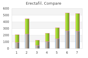
| Comparative prices of Erectafil | ||
| # | Retailer | Average price |
| 1 | H-E-B | 624 |
| 2 | Bon-Ton Stores | 638 |
| 3 | Williams-Sonoma | 502 |
| 4 | Menard | 805 |
| 5 | Family Dollar | 818 |

